Uveal melanoma
Cyberknife treatment in Munich
The Munich center has already treated a large number of patients with uveal melanoma and thus offers a comprehensive radiosurgical experience with this disease.
With the Cyberknife technology, uveal melanoma is treated on an outpatient basis without surgical intervention. There are no complications due to fixation or open surgery. Inpatient hospitalization is not required. Likewise, no follow-up treatment or rehabilitation stay is required. For patients, this means they can keep their usual routine. The usual activities can be followed up in most cases following the treatment.
The treatment indication is provided in close consultation with the colleagues of the Eye Clinic in the Mathildenstraße of the University of Munich LMU. In individual cases, it is decided whether a Cyberknife treatment is the best and safest therapy option.
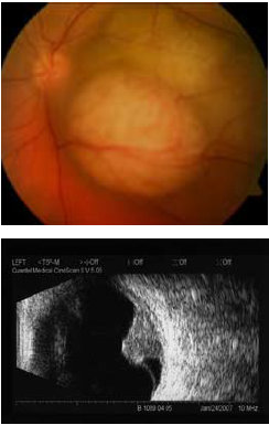
nach der Cyberknife-Behandlung.
Im Ultraschallbild sieht man nur
noch eine kleine Narbe im Vergleich
zu vorher.
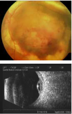
The uveal melanoma is an initially flat-growing tumor that progressively bulges, lifting the retina above it. The usually unilateral occurring tumor can form at different points in the eye, including the posterior pole. The annual incidence rate is 7 patients out of 1 million people.
In the beginning, the patients usually have no complaints. As the tumor size increases, the tumor becomes noticeable due to reduced visual performance or a shadow in the visual field. Rarely, eye pressure increase or breakthrough of the tumor through the dermis can be observed.
For examination and diagnosis, the eye is first examined by direct and indirect ophthalmology. In addition, an ultrasound sonography is performed to clarify tumor height (prominence), the internal echo (reflectivity), the presence of vessels and a retinal detachment (exudative ablatio retinae). With angiography of the fundus, tumorous vessels can be visualized. At diagnosis, only 1 percent of patients have metastases. These are often found in the liver.
For patients with uveal melanoma there are different therapies such as radiotherapy, radiosurgery and surgery.
Modern technologies include cyberknife radiosurgery. This is done without fixing the patient, the eye is immobilized by retrobulbar anesthesia. The radiosurgical cyberknife method can be used to destroy not only small but also medium to large melanomas without surgical intervention. The experienced Cyberknife Zentrum München offers its patients safe therapy.
Other radiotherapeutic and radiosurgical methods of treatment are also used. These include brachytherapy with the application of radioactive radiation carriers (Ruthenium 106 plaques) to the dermis of the eye, conventional Linac radiation therapy, proton therapy and the older gamma-knife method.
A previously widespread treatment of choroidal melanoma was the surgical removal of the eye (enucleation), especially in large initial findings with the aim to prevent metastasis of the tumor. However, large studies have failed to establish a link between different primary tumor treatments and the later onset of metastases.


Behandlungsanfragen
Please use our contact button for a treatment inquiry and individual appointment.
Contact